Home > Arts > Artists > J > Jacob Jacobs
Hand muscle anatomy, 1831 artwork

![]()
Wall Art and Photo Gifts from Science Photo Library
Hand muscle anatomy, 1831 artwork
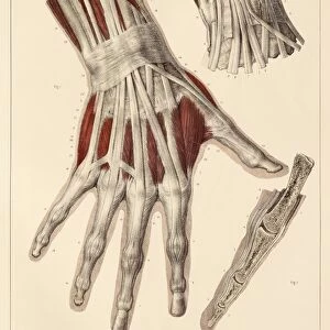
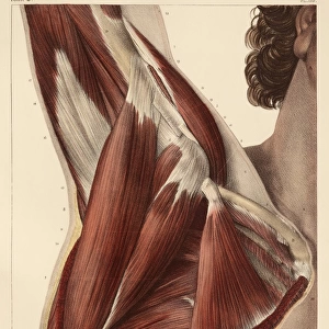
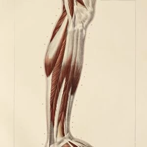
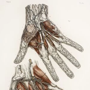
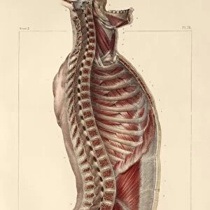
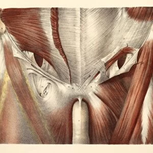
Hand muscle anatomy. Radial view of the ligaments and muscles of the hand (main artwork, centre). The insets show various aspects of the ligaments, muscles, and bones of the fingers. This anatomical artwork is plate 123 from volume 2 (1831) of Traite complet de l anatomie de l homme (1831-1854). This 8-volume anatomy atlas was produced by the French physician and anatomist Jean-Baptiste Marc Bourgery (1797-1849). The illustrations were by Nicolas-Henri Jacob (1781-1871)
Science Photo Library features Science and Medical images including photos and illustrations
Media ID 9269847
© SCIENCE PHOTO LIBRARY
1831 Anatomical Artwork Anatomical Illustration Anatomy Atlas Bones Finger Fingers French Hand Inset Insets Jean Baptiste Marc Bourgery Lateral Ligament Ligaments Muscles Nicolas Henri Jacob Phalanges Phalanx Volume 2 Volume Ii Wrist Musculature
FEATURES IN THESE COLLECTIONS
> Arts
> Artists
> J
> Jacob Jacobs
EDITORS COMMENTS
This print showcases the intricate hand muscle anatomy as depicted in an 1831 artwork. The central image portrays a radial view of the ligaments and muscles that compose the human hand, serving as a testament to the remarkable complexity of this vital body part. Surrounding insets provide further insight into various aspects of finger ligaments, muscles, and bones. The artwork originates from plate 123 in volume 2 (1831) of "Traite complet de l'anatomie de l'homme" an esteemed eight-volume anatomy atlas created by Jean-Baptiste Marc Bourgery, a renowned French physician and anatomist. The illustrations themselves were skillfully crafted by Nicolas-Henri Jacob. This historical piece offers both educational value and aesthetic appeal. Its detailed portrayal allows viewers to appreciate the musculature and ligamentous structures that enable our hands to perform countless tasks with precision and dexterity. From the phalanges to the wrist, every element is meticulously rendered for thorough examination. As we delve into this mesmerizing image, we are reminded of how far medical knowledge has advanced since its creation over two centuries ago. Yet it also serves as a timeless reminder of our shared humanity—regardless of time period or nationality—as we all possess these intricate anatomical features within us. This print from Science Photo Library provides a fascinating glimpse into medical history while celebrating the beauty and complexity inherent in our own bodies.
MADE IN THE USA
Safe Shipping with 30 Day Money Back Guarantee
FREE PERSONALISATION*
We are proud to offer a range of customisation features including Personalised Captions, Color Filters and Picture Zoom Tools
SECURE PAYMENTS
We happily accept a wide range of payment options so you can pay for the things you need in the way that is most convenient for you
* Options may vary by product and licensing agreement. Zoomed Pictures can be adjusted in the Cart.