Home > Science > SEM
Rod and cone cells of the eye, SEM C014 / 4866

![]()
Wall Art and Photo Gifts from Science Photo Library
Rod and cone cells of the eye, SEM C014 / 4866
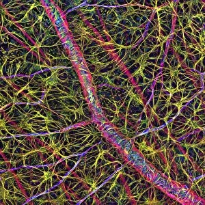
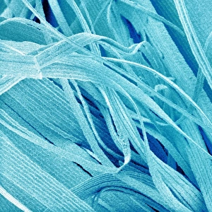
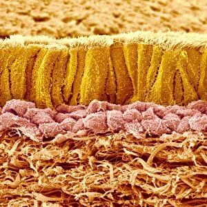
Rod and cone cells of the eye. Coloured scanning electron micrograph (SEM) of rod and cone cells in the retina of a mammalian eye. Cone cells and the more numerous rod cells are specialised light-sensitive cells that occur on the surface of the retina. They are responsible for detecting visible images, which are transmitted as nerve impulses to the optic nerve and the brain. There are about 130 million rod cells in the human retina, which detect light intensity and so are important for day and night vision. The less numerous cone-like cone cells (about 6.5 million in the human retina) respond specifically to colour. Magnification: x5, 300 when printed at 10 centimetres wide
Science Photo Library features Science and Medical images including photos and illustrations
Media ID 9224795
© CLOUDS HILL IMAGING LTD/SCIENCE PHOTO LIBRARY
Colored Cone Cell Cones Light Sensitive Mammal Mammalian Neural Neuron Neuronal Photoreceptor Photoreceptors Receptor Receptors Reptile Retina Retinal Rods Sense Sensory Sight Vision Visual Cells Nervous System
EDITORS COMMENTS
This print showcases the intricate world of rod and cone cells in the eye. In this colored scanning electron micrograph (SEM), we are granted a glimpse into the retina of a mammalian eye, where these specialized light-sensitive cells reside. The rod and cone cells play a vital role in our visual perception by detecting visible images and transmitting them as nerve impulses to the optic nerve and ultimately, to our brain. The human retina houses an impressive 130 million rod cells that excel at detecting light intensity, making them essential for both day and night vision. Alongside these rods, we find approximately 6.5 million cone-like cone cells that respond specifically to color stimuli. Together, they form an intricate network responsible for capturing the essence of what we see. With a magnification of x5,300 when printed at 10 centimeters wide, this image allows us to appreciate the remarkable detail present within these tiny sensory receptors. Each cell is meticulously structured to fulfill its unique function within our visual system. As we delve into this mesmerizing microcosm captured by Clouds Hill Imaging Ltd/Science Photo Library through their state-of-the-art scanning electron microscope (SEM), it becomes evident how nature has intricately designed every aspect of our eyesight. This photograph serves as a testament to both the beauty and complexity found within our own anatomy while reminding us of the wonders that lie just beyond what meets the naked eye.
MADE IN THE USA
Safe Shipping with 30 Day Money Back Guarantee
FREE PERSONALISATION*
We are proud to offer a range of customisation features including Personalised Captions, Color Filters and Picture Zoom Tools
SECURE PAYMENTS
We happily accept a wide range of payment options so you can pay for the things you need in the way that is most convenient for you
* Options may vary by product and licensing agreement. Zoomed Pictures can be adjusted in the Cart.