White blood cell antigen presentation C016 / 9058

![]()
Wall Art and Photo Gifts from Science Photo Library
White blood cell antigen presentation C016 / 9058
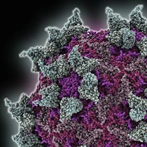
White blood cell antigen presentation. Coloured scanning electron micrograph (SEM) showing the interaction between a macrophage (yellow) and a T helper lymphocyte (Th cell, green), two components of the bodys immune system. Both are types of white blood cell. Macrophages are antigen-presenting cells (APCs). They present antigens (fragments on the surface of pathogens or foreign objects) to T lymphocytes, activating them. Each T lymphocyte recognises and binds to a specific antigen. Binding of the Th cell to the antigen presented by the macrophage activates the Th cell. This leads to its proliferation and the activation of other immune cells that eliminate the antigen. Magnification: x8000 when printed 10cm wide
Science Photo Library features Science and Medical images including photos and illustrations
Media ID 9246161
© STEVE GSCHMEISSNER/SCIENCE PHOTO LIBRARY
Activating Activation Binding Cell Biology Colored Cytological Cytology Electron Microscope Haematological Haematology Hematological Hematology Immune System Immunity Immunological Immunology Interacting Interaction Leucocyte Leucocytes Leukocyte Leukocytes Macrophage Pathogenic Presenting Recognising Recognition T Lymphocyte White Blood Cell Cells Pathogen
EDITORS COMMENTS
This print titled "White Blood Cell Antigen Presentation" offers a mesmerizing glimpse into the intricate workings of our body's immune system. In this colored scanning electron micrograph (SEM), we witness the dynamic interaction between a macrophage and a T helper lymphocyte, both essential components of our white blood cells. The macrophage, depicted in vibrant yellow, acts as an antigen-presenting cell (APC). Its crucial role is to present antigens - fragments found on the surface of pathogens or foreign objects - to T lymphocytes. These antigens serve as recognition markers for specific pathogens that invade our bodies. In striking green, the T helper lymphocyte binds itself to the antigen presented by the macrophage, effectively activating it. This activation leads to proliferation and triggers other immune cells' response to eliminate the harmful antigen. At a magnification of x8000 when printed 10cm wide, this image showcases remarkable detail and precision in capturing this critical biological process. It highlights how these microscopic interactions play a vital role in safeguarding our health against pathogenic invaders. Photographed by Steve Gschmeissner from Science Photo Library, this image seamlessly combines artistry with scientific inquiry. It serves as a testament to humanity's ongoing quest for understanding and harnessing the power of our immune system for medical advancements and improved well-being.
MADE IN THE USA
Safe Shipping with 30 Day Money Back Guarantee
FREE PERSONALISATION*
We are proud to offer a range of customisation features including Personalised Captions, Color Filters and Picture Zoom Tools
SECURE PAYMENTS
We happily accept a wide range of payment options so you can pay for the things you need in the way that is most convenient for you
* Options may vary by product and licensing agreement. Zoomed Pictures can be adjusted in the Cart.