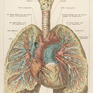
Liver and stomach arteries, 1825 artwork

![]()
Wall Art and Photo Gifts from Science Photo Library
Liver and stomach arteries, 1825 artwork
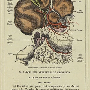
Liver and stomach arteries. Dissection of the abdomen, showing the arteries (red) of the liver (brown, upper left), stomach (pink, upper centre), and the intestines (grey with yellow fat, lower centre). The gall bladder (green), bile duct (also green), and the portal vein (blue) are also shown. This anatomical artwork is plate 228 from volume 4 of Manuel d anatomie descriptive du corps humain (1825). This 5-volume anatomy atlas was produced by French physician and surgeon Jules Germain Cloquet (1790-1883). The illustrations were by Haincelin. Volume 4 illustrated the anatomy of the circulatory and respiratory systems
Science Photo Library features Science and Medical images including photos and illustrations
Media ID 9223295
© SCIENCE PHOTO LIBRARY
1825 Abdomen Abdominal Anatomical Artwork Anatomical Illustration Anatomy Atlas Anterior Arterial System Arteries Blood Vessels Digestive System Dissected Dissection French Frontal Gall Bladder Gastric Gastrointestinal Haincelin Hepatic Intestinal Intestine Intestines Jules Germain Cloquet Liver Oxygenated Blood Portal Vein Stomach Vascular Volume 4 Volume Iv Artery Blood Vessel Circulatory System
EDITORS COMMENTS
This artwork, titled "Liver and Stomach Arteries" offers a remarkable glimpse into the intricate anatomy of the human body. Created in 1825 by French physician and surgeon Jules Germain Cloquet, this illustration is part of his renowned five-volume atlas on human anatomy. Plate 228 from volume 4 specifically focuses on the circulatory and respiratory systems. The detailed dissection showcased in this print reveals the vibrant red arteries that supply oxygenated blood to both the liver and stomach. The brownish hue represents the liver, positioned prominently in the upper left corner, while the pink area signifies the stomach located at the upper center. Below them lies a depiction of grey intestines adorned with yellow fat. Noteworthy elements such as the gall bladder (green), bile duct (also green), and portal vein (blue) are also meticulously depicted within this artwork. These components play crucial roles in digestion and circulation within our bodies. The historical significance of this piece cannot be overstated; it provides invaluable insight into medical practices during early 19th-century France. Crafted with precision by artist Haincelin under Cloquet's guidance, this anatomical illustration exemplifies their commitment to advancing scientific knowledge. Whether you're an enthusiast of medical history or simply fascinated by human anatomy, this print serves as a testament to our ongoing quest for understanding our own bodies' intricacies.
MADE IN THE USA
Safe Shipping with 30 Day Money Back Guarantee
FREE PERSONALISATION*
We are proud to offer a range of customisation features including Personalised Captions, Color Filters and Picture Zoom Tools
SECURE PAYMENTS
We happily accept a wide range of payment options so you can pay for the things you need in the way that is most convenient for you
* Options may vary by product and licensing agreement. Zoomed Pictures can be adjusted in the Cart.