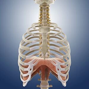
Diaphragm anatomy, 1831 artwork

![]()
Wall Art and Photo Gifts from Science Photo Library
Diaphragm anatomy, 1831 artwork
Diaphragm anatomy, posterior view. The diaphragm is the white area at top, connected to surrounding muscles (red). Part of the spine is at bottom, and ribs at left and right. The relaxation and upwards movement of the diaphragm helps the lungs to deflate during respiration. This anatomical artwork is plate 78 from volume 2 (1831) of Traite complet de l anatomie de l homme (1831-1854). This 8-volume anatomy atlas was produced by the French physician and anatomist Jean-Baptiste Marc Bourgery (1797-1849). The illustrations were by Nicolas-Henri Jacob (1781-1871)
Science Photo Library features Science and Medical images including photos and illustrations
Media ID 9273385
© SCIENCE PHOTO LIBRARY
1831 Abdomen Abdominal Anatomical Artwork Anatomical Illustration Anatomy Atlas Backbone Chest Diaphragm Dissected French From Behind Internal Jean Baptiste Marc Bourgery Muscles Nicolas Henri Jacob Posterior View Ribs Thoracic Thorax Torso Trunk Volume 2 Volume Ii Musculature
EDITORS COMMENTS
This print showcases a detailed artwork of the diaphragm anatomy from 1831. In this posterior view, the diaphragm is depicted as a white area at the top, intricately connected to surrounding muscles highlighted in red. The lower part of the spine and ribs are also visible on the left and right sides respectively. This anatomical masterpiece, plate 78 from volume 2 of Traite complet de l'anatomie de l'homme (1831-1854), was created by renowned French physician and anatomist Jean-Baptiste Marc Bourgery (1797-1849). The illustrations were skillfully crafted by Nicolas-Henri Jacob (1781-1871). The diaphragm plays a vital role in respiration by aiding in both relaxation and upward movement, facilitating lung deflation during breathing. This historical artwork offers an intriguing glimpse into early medical knowledge and serves as a testament to Bourgery's contributions to human anatomy. With its intricate detailing and accurate representation of internal structures, this image provides valuable insights into abdominal musculature while highlighting the complexity of our own bodies. It serves as a reminder of how far we have come in understanding human physiology since its creation over two centuries ago. This remarkable piece captures not only scientific significance but also artistic beauty, making it an invaluable addition to any collection or display dedicated to medical history or anatomical studies.
MADE IN THE USA
Safe Shipping with 30 Day Money Back Guarantee
FREE PERSONALISATION*
We are proud to offer a range of customisation features including Personalised Captions, Color Filters and Picture Zoom Tools
SECURE PAYMENTS
We happily accept a wide range of payment options so you can pay for the things you need in the way that is most convenient for you
* Options may vary by product and licensing agreement. Zoomed Pictures can be adjusted in the Cart.